Home
/ Anatomy Of Right Side Of Back Of Rib Cage - Dr Gaytri Gandotra Rib Cage Kidney Shoulder Vertebral Column Kidney Abdomen Organ Png Pngegg - To better understand slipping rib syndrome and how it may develop, a quick review of the related anatomy is needed.
Anatomy Of Right Side Of Back Of Rib Cage - Dr Gaytri Gandotra Rib Cage Kidney Shoulder Vertebral Column Kidney Abdomen Organ Png Pngegg - To better understand slipping rib syndrome and how it may develop, a quick review of the related anatomy is needed.
Anatomy Of Right Side Of Back Of Rib Cage - Dr Gaytri Gandotra Rib Cage Kidney Shoulder Vertebral Column Kidney Abdomen Organ Png Pngegg - To better understand slipping rib syndrome and how it may develop, a quick review of the related anatomy is needed.. It then hooks back to reach the right side with its relationship to the esophagus variable: When you reach the midpoint between the forelegs, make another incision down to the pan. Jun 10, 2021 · the thoracic cage (rib cage) is the skeleton of the thoracic wall. Key anatomical structures of the human body's rib cage related to slipped rib are illustrated. There are 12 pairs of ribs, and the first 10 articulate with both the thoracic spine and the costal cartilage on the front of the rib cage.
To better understand slipping rib syndrome and how it may develop, a quick review of the related anatomy is needed. If you follow the path of your ribs around from the front or sides of the back, you can feel where they attach to the thoracic vertebrae in the back. A short summary of this paper. When you reach the midpoint between the forelegs, make another incision down to the pan. It is supported by the vertical sternum or.
What Causes Pain Around The Ribs And Back Symptoms Regenexx from regenexx.com It is formed by the 12 thoracic vertebrae, 12 pairs of ribs and associated costal cartilages and the sternum. To better understand slipping rib syndrome and how it may develop, a quick review of the related anatomy is needed. Slipping rib syndrome's connection to the thoracic spine and pain. It is supported by the vertical sternum or. A short summary of this paper. The diaphragm should remain intact, but now the rib cage can be pulled back and pinned to the pan, exposing the thoracic cavity. If you follow the path of your ribs around from the front or sides of the back, you can feel where they attach to the thoracic vertebrae in the back. 6 full pdfs related to this paper.
15% between esophagus and trachea
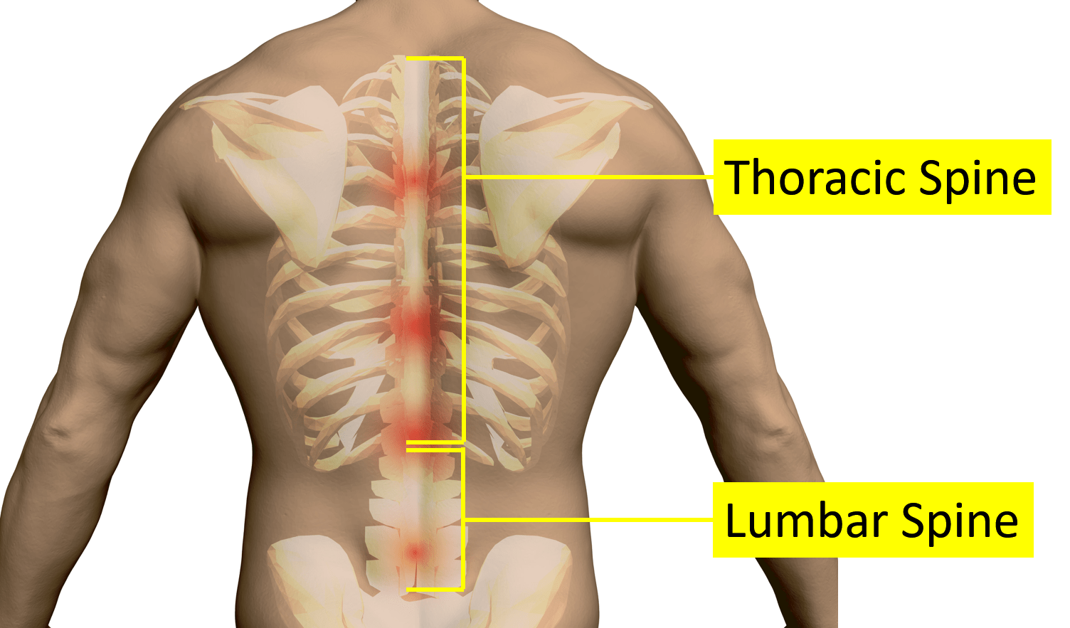
The remaining ribs are called floating ribs and only attach to the spine. Since the ribs and rib cage are attached to the spine, dysfunction in the spine can cause symptoms and problems in the ribs. The main function of the thoracic spine is to hold the rib cage which protects the heart and lungs. Instead of being the first branch (with the right common carotid as the brachiocephalic artery), it arises on its own as the fourth branch, distal to the left subclavian artery. It is supported by the vertical sternum or. 15% between esophagus and trachea The thoracic cage takes the form of a domed bird cage with the horizontal bars formed by ribs and costal cartilages. Jun 10, 2021 · the thoracic cage (rib cage) is the skeleton of the thoracic wall. It then hooks back to reach the right side with its relationship to the esophagus variable: Go back to the diaphragm area and use a scalpel to cut the wall of the body cavity away from the diaphragm. A short summary of this paper. 80% posterior to the esophagus; When you reach the midpoint between the forelegs, make another incision down to the pan.
Instead of being the first branch (with the right common carotid as the brachiocephalic artery), it arises on its own as the fourth branch, distal to the left subclavian artery. To better understand slipping rib syndrome and how it may develop, a quick review of the related anatomy is needed. It is supported by the vertical sternum or. Slipping rib syndrome's connection to the thoracic spine and pain. It then hooks back to reach the right side with its relationship to the esophagus variable:
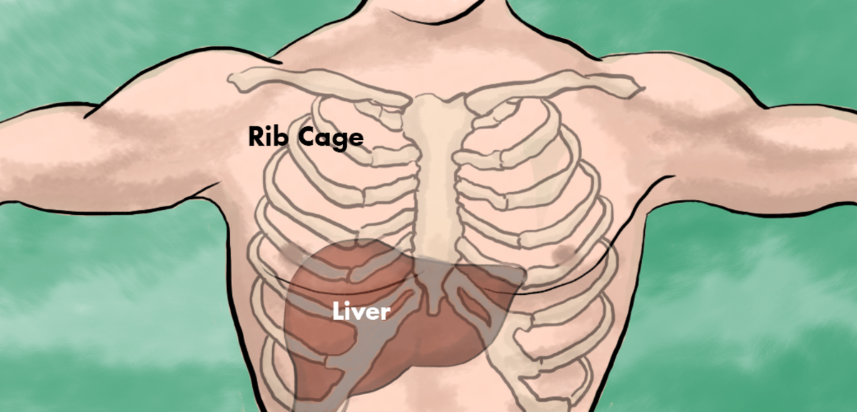
Has The Liver Always Been Completely Behind The Rib Cage Sherdog Forums Ufc Mma Boxing Discussion from usercontent2.hubstatic.com It is formed by the 12 thoracic vertebrae, 12 pairs of ribs and associated costal cartilages and the sternum. 15% between esophagus and trachea There are 12 pairs of ribs, and the first 10 articulate with both the thoracic spine and the costal cartilage on the front of the rib cage. 80% posterior to the esophagus; Jun 10, 2021 · the thoracic cage (rib cage) is the skeleton of the thoracic wall. The main function of the thoracic spine is to hold the rib cage which protects the heart and lungs. To better understand slipping rib syndrome and how it may develop, a quick review of the related anatomy is needed. Instead of being the first branch (with the right common carotid as the brachiocephalic artery), it arises on its own as the fourth branch, distal to the left subclavian artery.
15% between esophagus and trachea
A short summary of this paper. Go back to the diaphragm area and use a scalpel to cut the wall of the body cavity away from the diaphragm. Since the ribs and rib cage are attached to the spine, dysfunction in the spine can cause symptoms and problems in the ribs. 15% between esophagus and trachea It then hooks back to reach the right side with its relationship to the esophagus variable: The thoracic cage takes the form of a domed bird cage with the horizontal bars formed by ribs and costal cartilages. Instead of being the first branch (with the right common carotid as the brachiocephalic artery), it arises on its own as the fourth branch, distal to the left subclavian artery. The main function of the thoracic spine is to hold the rib cage which protects the heart and lungs. It is formed by the 12 thoracic vertebrae, 12 pairs of ribs and associated costal cartilages and the sternum. There are 12 pairs of ribs, and the first 10 articulate with both the thoracic spine and the costal cartilage on the front of the rib cage. Jul 08, 2019 · thoracic spine (mid back) the twelve thoracic vertebrae, t1 to t12, are connected to your ribs. To better understand slipping rib syndrome and how it may develop, a quick review of the related anatomy is needed. 6 full pdfs related to this paper.
Jun 10, 2021 · the thoracic cage (rib cage) is the skeleton of the thoracic wall. If you follow the path of your ribs around from the front or sides of the back, you can feel where they attach to the thoracic vertebrae in the back. It then hooks back to reach the right side with its relationship to the esophagus variable: The diaphragm should remain intact, but now the rib cage can be pulled back and pinned to the pan, exposing the thoracic cavity. When you reach the midpoint between the forelegs, make another incision down to the pan.
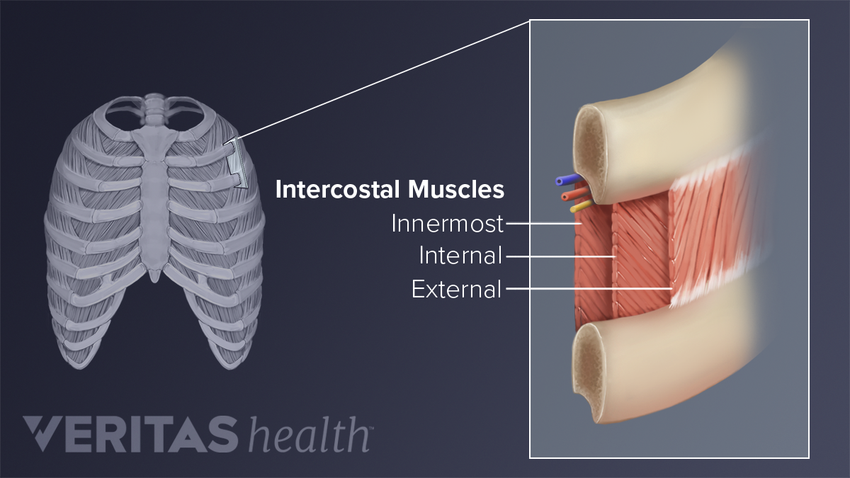
Upper Back Pain From Intercostal Muscle Strain from embed.widencdn.net When you reach the midpoint between the forelegs, make another incision down to the pan. It is formed by the 12 thoracic vertebrae, 12 pairs of ribs and associated costal cartilages and the sternum. 15% between esophagus and trachea 80% posterior to the esophagus; Go back to the diaphragm area and use a scalpel to cut the wall of the body cavity away from the diaphragm. 6 full pdfs related to this paper. Since the ribs and rib cage are attached to the spine, dysfunction in the spine can cause symptoms and problems in the ribs. To better understand slipping rib syndrome and how it may develop, a quick review of the related anatomy is needed.
A short summary of this paper.
If you follow the path of your ribs around from the front or sides of the back, you can feel where they attach to the thoracic vertebrae in the back. Go back to the diaphragm area and use a scalpel to cut the wall of the body cavity away from the diaphragm. A short summary of this paper. Since the ribs and rib cage are attached to the spine, dysfunction in the spine can cause symptoms and problems in the ribs. Jun 10, 2021 · the thoracic cage (rib cage) is the skeleton of the thoracic wall. It is supported by the vertical sternum or. Instead of being the first branch (with the right common carotid as the brachiocephalic artery), it arises on its own as the fourth branch, distal to the left subclavian artery. The main function of the thoracic spine is to hold the rib cage which protects the heart and lungs. There are 12 pairs of ribs, and the first 10 articulate with both the thoracic spine and the costal cartilage on the front of the rib cage. It is formed by the 12 thoracic vertebrae, 12 pairs of ribs and associated costal cartilages and the sternum. Key anatomical structures of the human body's rib cage related to slipped rib are illustrated. 6 full pdfs related to this paper. When you reach the midpoint between the forelegs, make another incision down to the pan.